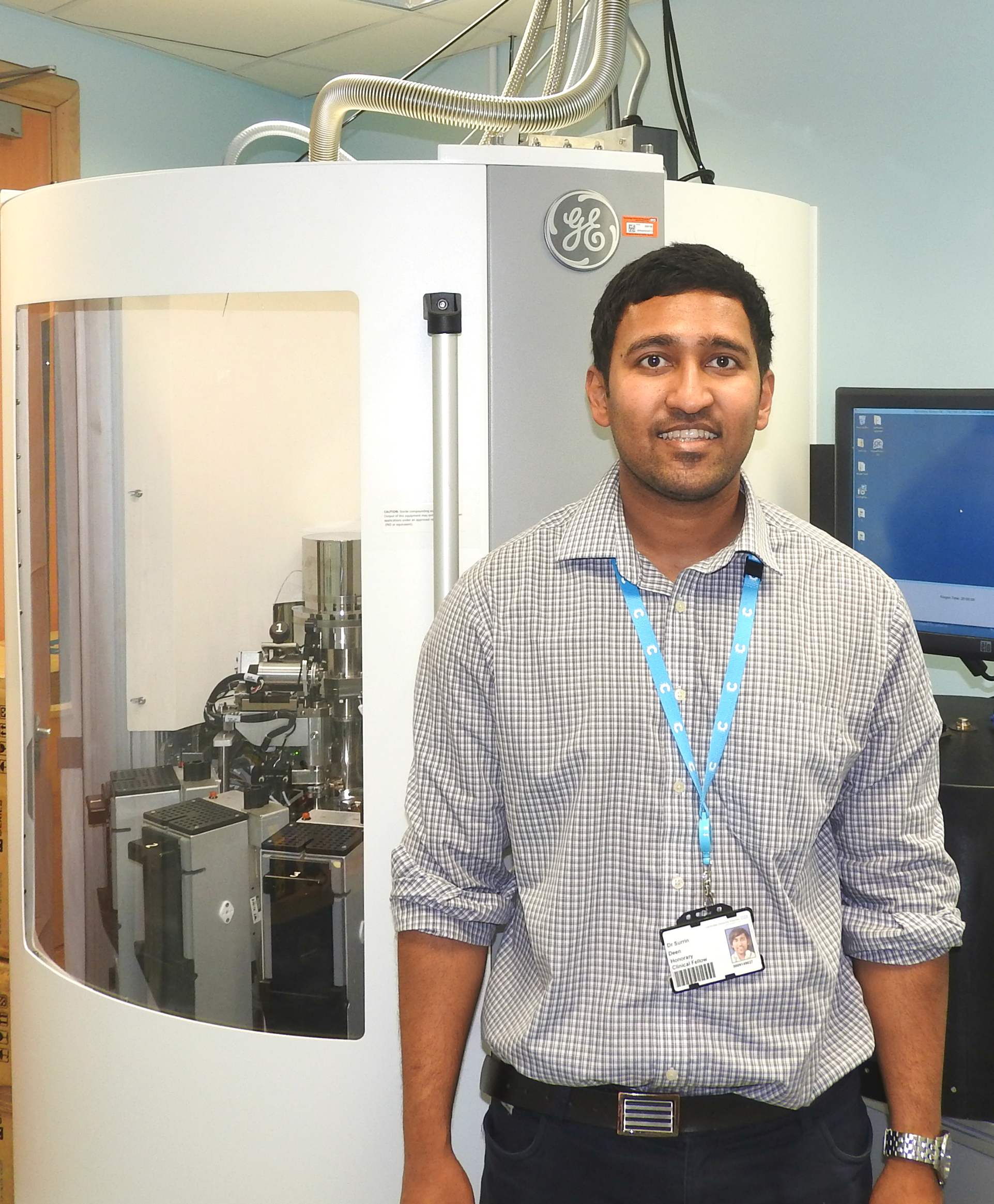
Surrin Deen speaks about his research into hyperpolarized MRI and its importance for the early detection of cancer.
Individual patients respond differently to the same treatment and hyperpolarized imaging can help physicians provide more personalised care and avoid treatment side effects.
Surrin Deen
It’s not every PhD researcher who becomes involved in a race to be the first in Europe to use a new technique which could have a major impact on healthcare, but Surrin Deen’s work on hyperpolarized MRI – a new, more detailed MRI scan – provided just that experience.
Hyperpolarized MRI can increase scientists' ability to detect the breakdown products of glucose in cancer by more than 10,000 fold. Surrin says it can diagnose very early stage cancers, improving treatment outcomes, and also help scientists to better understand the biology of cancer and other diseases.
Over the course of his PhD, Surrin’s team overcame challenges like securing a reliable supply of carbon 13-labelled molecules and developing methods to ensure that the sterility of the molecules was not compromised during human imaging. A large initial part of his PhD was dedicated to designing the research study protocol and applying for ethical approval to inject and image carbon 13-labelled molecules in humans.
Surrin’s mixed background in medicine and physics helped him to link the multidisciplinary technological and medical aspects of the project. “My role was to co-ordinate the efforts of a large team of physicists, engineers, oncologists and surgeons to ensure we could test the imaging process on humans as early on as possible,” he says.
Once the equipment was ready, there was a race to be the first team to use it successfully on humans to detect cancer. In March 2016 Cambridge became the first site outside of North America to successfully image a human with hyperpolarized carbon-13 MRI.
Surrin’s PhD is now focusing on the application of hyperpolarized MRI – as well as sodium and diffusion MRI – to ovarian cancer. He says: "Individual patients respond differently to the same treatment and hyperpolarized imaging can help physicians provide more personalised care and avoid treatment side effects. For example, we can image a patient the day after the start of their chemotherapy to evaluate if the drug we have given is having any effect. Based on this information we can then either immediately switch to a more effective therapy or continue with the same treatment. Before hyperpolarized imaging this information would not have been available until weeks or months later meaning that a patient could be given an ineffective therapy for a long time without anyone realising it and during which time the cancer would continue to spread."
Background
From an early age Surrin [2014] has been keen to put his ability in Maths and Physics to practical use. Born in Trinidad and Tobago, Surrin excelled at Maths. During his mid-teens he represented his country at the International Mathematical Olympiads in Greece and Mexico. His early educational experiences made him realise the benefit that could come from applying Mathematics and Physics to healthcare and he felt that radiology was the speciality in medicine that could make the best use of technology to improve the lives of patients.
He was awarded a scholarship for undergraduate studies in medicine at King’s College London and also completed a one-year Bachelor’s Degree in Medical Physics and Bioengineering at University College London. Following his medical degree, Surrin worked in vascular surgery, interventional radiology and emergency medicine at Royal Oldham Hospital where he gained his unrestricted licence to practice medicine.
He then contacted his current supervisor Ferdia Gallagher about his interest in radiology and was introduced to the hyperpolarized MRI project that he began working on as a PhD student in 2014. Surrin is now in the process of writing up his PhD thesis and applying for jobs to further his career in radiology. He says he is keen to continue his involvement in research alongside working as a clinical radiologist.

Surrin Deen
- Alumni
- Trinidad and Tobago
- 2014 PhD Radiology
- Trinity Hall
Surrin is a medical doctor based at Addenbrooke's Hospital and Cancer Research UK, Cambridge Institute, with a background of undergraduate studies in Medical Physics and Bioengineering.
His research at Cambridge is translational and involves the use of MRI to image metabolism in ovarian and other cancers after the injection of tracers labelled with hyperpolarized nuclei that enhance detection by a factor of several thousand fold.
The imaging results are compared to histology and immunohistochemistry of cancerous tissue sampled by biopsy and at surgery to validate the findings of the imaging and to show that it is possible to detect cancers earlier and to non-invasively monitor the response of cancers to different treatments using hyperpolarized nuclei.


