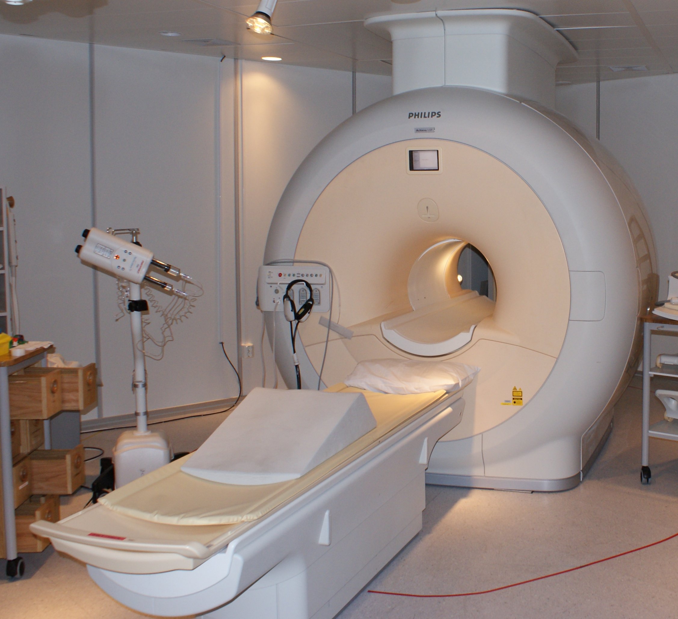
Surrin Deen takes part in first human liver scan using metabolic imaging system.
A Gates Cambridge Scholar is part of the team that performed the world's first hyperpolarized MRI image of the human liver – an achievement which could have huge implications for the early detection and successful treatment of cancer.
University of Cambridge researchers, including Gates Cambridge Scholar Surrin Deen [2014], are the first in Europe to use this type of MRI on humans as well as the first in the world to image a human liver.
Cancer is a major public health problem. In the developing world cancer causes more deaths than HIV/AIDS, tuberculosis and malaria combined. According to Cancer Research UK half of all people born in the UK after 1960 will develop cancer. This means the condition is likely to affect almost every household in the future.
Cancers consume large amounts of glucose to provide the energy they need to sustain their rapid growth. As a result breakdown products of glucose such as lactate build up within cancerous cells. This build-up occurs early on in the development of cancers, long before the tumours have grown large enough to cause symptoms or to become visible on conventional imaging.
Hyperpolarized MRI has recently been shown to be capable of increasing the ability to the detect breakdown products of glucose in cancer by more than 10,000 fold. Now this technology is being translated into the clinical setting for the first time where it is expected to completely change the prognosis of cancer by facilitating earlier diagnosis and treatment. Early treatment before cancer has spread is known to significantly improve outcome and survival.
There are a wide range of cancer therapies available and it is often a challenge to select the best option. One of the most promising applications of hyperpolarised MRI is in identifying a cancer’s response
to treatment. Animal studies at Cambridge have demonstrated that a tumour’s response to chemotherapy or radiotherapy can be detected on hyperpolarized imaging in as little as 24 hours after treatment.
Surrin, who is doing a PhD on the use of hyperpolarized MRI in ovarian cancer, says: "If the same applies to humans, hyperpolarized imaging can help physicians provide more personalised care and avoid treatment side effects. For example we could image a patient the day after the start of chemotherapy to evaluate if the drug we have given is having any effect. Based on this information we can then either immediately switch to a more effective therapy or continue with the same treatment. Before hyperpolarized imaging this information would not have been available until weeks or months later."
Surrin is in the second year of his PhD and is supervised in his work by Dr Ferdia Gallagher and Dr James Brenton. Hyperpolarized imaging research at Cambridge is also supported by Professor Kevin Brindle, one of the pioneers in the development of clinical hyperpolarization techniques. The research is funded by grants from the Wellcome Trust and Cancer Research UK.
*Picture credit: Traditional MRI scanning c/o Wikipedia.

Surrin Deen
- Alumni
- Trinidad and Tobago
- 2014 PhD Radiology
- Trinity Hall
Surrin is a medical doctor based at Addenbrooke's Hospital and Cancer Research UK, Cambridge Institute, with a background of undergraduate studies in Medical Physics and Bioengineering.
His research at Cambridge is translational and involves the use of MRI to image metabolism in ovarian and other cancers after the injection of tracers labelled with hyperpolarized nuclei that enhance detection by a factor of several thousand fold.
The imaging results are compared to histology and immunohistochemistry of cancerous tissue sampled by biopsy and at surgery to validate the findings of the imaging and to show that it is possible to detect cancers earlier and to non-invasively monitor the response of cancers to different treatments using hyperpolarized nuclei.


